The balancing electrons is employed by laboratory and hospital personnel for very precise measurement of chemical substances, powders, and living organisms. Its accuracy to the tune of balancing electrons allows making medications in exact doses, calibrating standards, and preparing diagnostic reagents. balancing electrons not only gives undistorted and sensitive measures but also improves lab operation, helps to control quality, and keeps clinical research intact. Its importance is appreciated in both hospital tests and medical product development as dependable.

balancing electrons are used in clinical laboratories for making calibration references that are then checked on the analytics machines. Exact weighing guarantees that the calibration materials keep the same mass values throughout the uses they are subjected to. This application endorses instrument accuracy checks, routine audits in laboratories, and compliance with government regulations. By being a part of trustworthy calibration workflows, balancing electrons is a major factor and, thus, a contributor to measurement accuracy kept in hospitals' diagnostic devices.

The era of balancing electrons in hospitals will go beyond the traditional settings and embrace multidisciplinary research environments. As the partnership of clinical, pharmaceutical, and biomedical teams becomes more robust, the analytical balances will cater to different experimental needs. By taking on various analytical actions, balancing electrons will still be a fundamental tool in contemporary hospital laboratory ecosystems.

balancing electrons in clinics is, however, maintained through regular performance verification conducted in the laboratories. Certified test weights are used to prove the reliability of the measurements with the passage of time. Verification activities being documented also implies good traceability and makes it easier for internal audits to take place. By making verification part of the routine maintenance, hospitals make sure that balancing electrons still gives trustworthy results for research and diagnostic workflows.
balancing electrons plays an important role in the hospital pharmacy in the accurate formulation of medications, intravenous solutions, and compounded drugs. Even slight alterations in the weight of drugs can change the effectiveness of the drug and endanger the patient's safety. The Pharmacy Technicians rely on balancing electrons for the correct dosing and checking of the active ingredients. The tool's accuracy guarantees the dependable preparation of drugs, the observance of the rules, and the quality control of the overall hospital pharmacy activities.
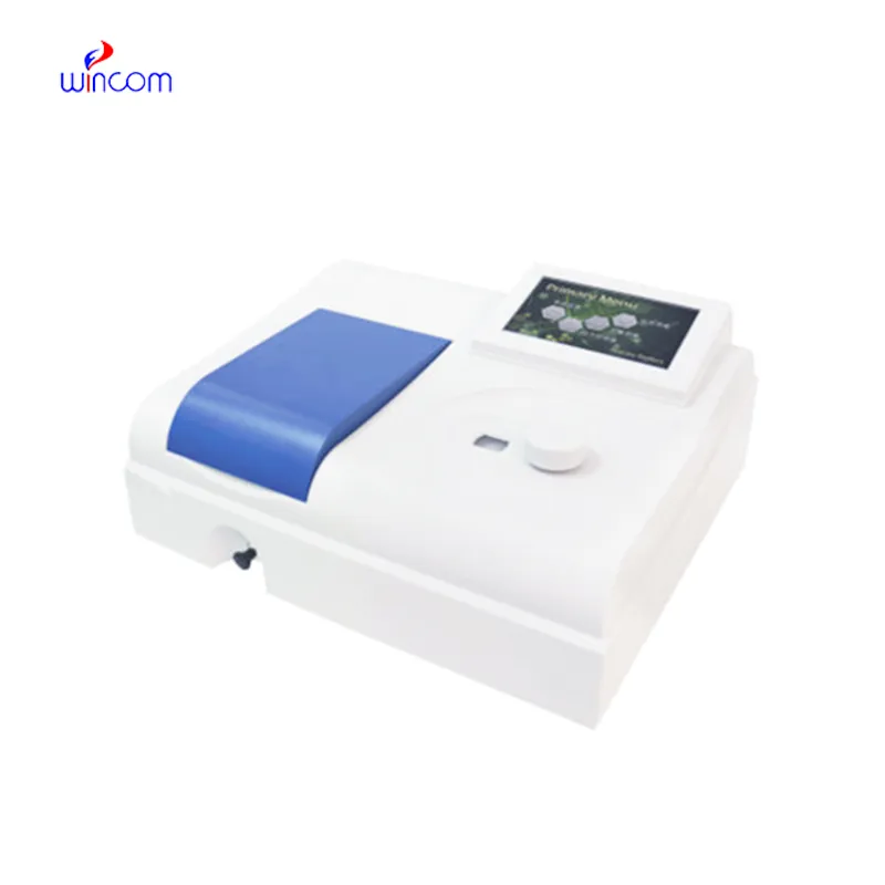
Q: What is the impact of temperature on the performance of analytical balance? A: The changes in temperature can lead to drift and weighing inconsistency. Q: Are analytical balances the only ones used in research laboratories? A: They are very important also for other processes such as sample preparation and improving the accuracy of the experiment. Q: How long does it usually take for an analytical balance to warm up? A: Warm-up times differ from one model to another, but an adequate stabilizing period increases the reliability of the measurement. Q: Is it possible for analytical balances to save weighing data? A: Internal memory or external data transfer are the two ways in which many models can achieve this feature. Q: Would it be necessary to undergo training if one wants to operate an analytical balance? A: Basic laboratory training will be enough to make sure that the balance is being used correctly.
The microscope delivers incredibly sharp images and precise focusing. It’s perfect for both professional lab work and educational use.
This x-ray machine is reliable and easy to operate. Our technicians appreciate how quickly it processes scans, saving valuable time during busy patient hours.
To protect the privacy of our buyers, only public service email domains like Gmail, Yahoo, and MSN will be displayed. Additionally, only a limited portion of the inquiry content will be shown.
Could you share the specifications and price for your hospital bed models? We’re looking for adjus...
We are planning to upgrade our imaging department and would like more information on your mri machin...
E-mail: [email protected]
Tel: +86-731-84176622
+86-731-84136655
Address: Rm.1507,Xinsancheng Plaza. No.58, Renmin Road(E),Changsha,Hunan,China

 af
af
 es
es
 ar
ar
 tr
tr
 sw
sw
 pt
pt
 th
th
 ur
ur
 bn
bn
 ne
ne
 vi
vi
 km
km
 lo
lo
 de
de
 ru
ru
 fi
fi
 nl
nl
 fa
fa
 fr
fr
 ko
ko