With the cutting-edge imaging processors, the fetal doppler monitor facilitates real-time, high-resolution images that are paramount in the detection of subtle physiological changes by clinicians. The display is user-friendly for easy parameter modification as well as image marking. The fetal doppler monitor exhibits the mix of effectiveness, mobility, and reliability for a huge range of diagnostic procedures.

In medical imaging, the fetal doppler monitor is a trustworthy device for different clinical departments. It provides vascular studies support by observing the state of the arteries and veins; in urology, it has a role in the assessment of the bladder and prostate health. The fetal doppler monitor also enables emergency medical staff to make quick trauma evaluations, directing their actions precisely and efficiently.

In addition, as new technologies emerge, the fetal doppler monitor is expected to become more compact and intelligent with better diagnostic capabilities. The new fetal doppler monitor will incorporate 3D and 4D capabilities. The fetal doppler monitor will also be integrated with digital hospitals for seamless management of patient data.

In order to extend the service life of the fetal doppler monitor, it is recommended that users refrain from applying much force during the process of connecting/disconnecting probes. Power cables should always remain dry. The fetal doppler monitor needs diagnostic tests to ensure that it produces quality images.
The fetal doppler monitor represents an advanced form of medical imaging technology that transforms sound waves into high-definition visual data. It is widely used for evaluating organ health, tracking fetal development, and detecting vascular conditions. The fetal doppler monitor ensures real-time monitoring and fast diagnostic results, supporting effective clinical workflows.
Q: What makes the ultrasound scannert effective for diagnostic imaging? A: Its high-frequency sound wave technology allows accurate visualization of internal body structures in real time. Q: How portable is the ultrasound scannert? A: The device features a compact and lightweight design, allowing easy movement between clinical departments. Q: What types of probes are compatible with the ultrasound scannert? A: It supports multiple probe types, including linear, convex, and phased array probes for varied diagnostic needs. Q: Does the ultrasound scannert require special training to operate? A: Basic technical training is recommended to maximize its imaging performance and functionality. Q: How long can the ultrasound scannert operate continuously? A: It is designed for extended use with efficient cooling systems and stable power performance.
The centrifuge operates quietly and efficiently. It’s compact but surprisingly powerful, making it perfect for daily lab use.
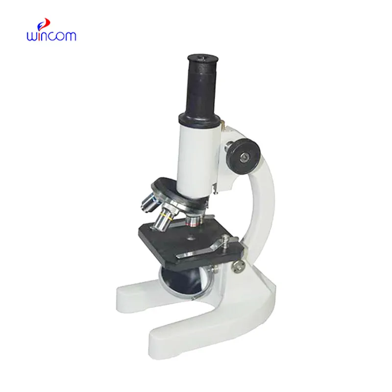
The microscope delivers incredibly sharp images and precise focusing. It’s perfect for both professional lab work and educational use.
To protect the privacy of our buyers, only public service email domains like Gmail, Yahoo, and MSN will be displayed. Additionally, only a limited portion of the inquiry content will be shown.
Could you share the specifications and price for your hospital bed models? We’re looking for adjus...
Hello, I’m interested in your water bath for laboratory applications. Can you confirm the temperat...
E-mail: [email protected]
Tel: +86-731-84176622
+86-731-84136655
Address: Rm.1507,Xinsancheng Plaza. No.58, Renmin Road(E),Changsha,Hunan,China

 af
af
 es
es
 ar
ar
 tr
tr
 sw
sw
 pt
pt
 th
th
 ur
ur
 bn
bn
 ne
ne
 vi
vi
 km
km
 lo
lo
 de
de
 ru
ru
 fi
fi
 nl
nl
 fa
fa
 fr
fr
 ko
ko