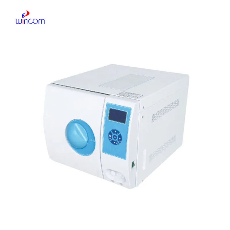
When it comes to durability and precision, the fetal heart doppler is a tough and highly accurate device that relies on the integration of the latest imaging software to provide quality diagnostic images. Its light structure and an electric system powered by rechargeable batteries make it a device that can be used at a stationary place or be taken out for mobile operations. The fetal heart doppler is a product that brings about a better workflow due to its easy-to-use controls and proper data management.

The fetal heart doppler is recognized for its great contribution to the field of surgery and thus is employed frequently in operating theaters for providing intraoperative guidance and ascertaining anatomical targets. It can easily locate areas where fluid has collected, determine the condition of the tissue, and provide evidence that the procedure has been successful. The fetal heart doppler can also be used dynamically and thus in sports medicine for imaging of muscles and tendons during movement analysis.

The fetal heart doppler should integrate with intelligent diagnostic ecosystems and communicate effortlessly with smartphones and electronic records. The synchronized exchange of data in real-time should enable constant patient observation. The next version should focus on improved design, better processing power of artificial intelligence algorithms, and enhanced reconstruction functions.

In order to retain the accuracy of the fetal heart doppler, it is important for operators to check the cables and connections of the transducers for evidence of wear. After each use, the surfaces should be wiped clean using non-abrasive cleaners. The fetal heart doppler should be turned off properly and covered to prevent dust from collecting. Regular checks by trained personnel should be done.
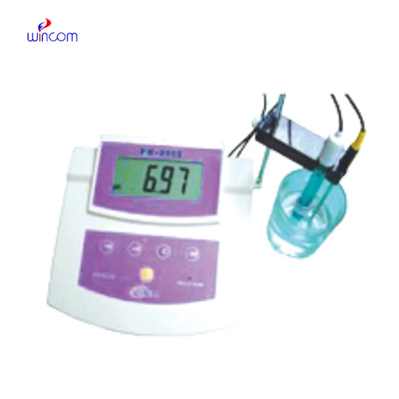
With the advanced imaging technology, the fetal heart doppler provides physicians unobstructed and precise images of internal organs. It is employed in the early disease diagnosis as well as in patient tracking. The fetal heart doppler functions by utilizing sound wave reflections to generate dynamic images, qualifying it as an essential tool in modern medical diagnostics. Through the fetal heart doppler, fast, non-invasive testing is facilitated for real-time assessment to support clinical decisions.
Q: What makes the ultrasound scannert effective for diagnostic imaging? A: Its high-frequency sound wave technology allows accurate visualization of internal body structures in real time. Q: How portable is the ultrasound scannert? A: The device features a compact and lightweight design, allowing easy movement between clinical departments. Q: What types of probes are compatible with the ultrasound scannert? A: It supports multiple probe types, including linear, convex, and phased array probes for varied diagnostic needs. Q: Does the ultrasound scannert require special training to operate? A: Basic technical training is recommended to maximize its imaging performance and functionality. Q: How long can the ultrasound scannert operate continuously? A: It is designed for extended use with efficient cooling systems and stable power performance.
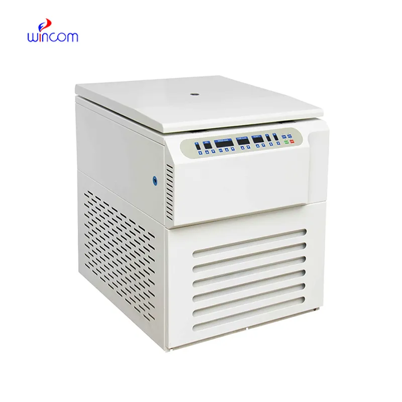
We’ve used this centrifuge for several months now, and it has performed consistently well. The speed control and balance are excellent.
The centrifuge operates quietly and efficiently. It’s compact but surprisingly powerful, making it perfect for daily lab use.
To protect the privacy of our buyers, only public service email domains like Gmail, Yahoo, and MSN will be displayed. Additionally, only a limited portion of the inquiry content will be shown.
Could you share the specifications and price for your hospital bed models? We’re looking for adjus...
I’d like to inquire about your x-ray machine models. Could you provide the technical datasheet, wa...
E-mail: [email protected]
Tel: +86-731-84176622
+86-731-84136655
Address: Rm.1507,Xinsancheng Plaza. No.58, Renmin Road(E),Changsha,Hunan,China

 af
af
 es
es
 ar
ar
 tr
tr
 sw
sw
 pt
pt
 th
th
 ur
ur
 bn
bn
 ne
ne
 vi
vi
 km
km
 lo
lo
 de
de
 ru
ru
 fi
fi
 nl
nl
 fa
fa
 fr
fr
 ko
ko