The hand held doppler fetal monitor has been designed considering the needs of modern health care, providing uninterrupted performance with its rapid image acquisition and high-definition visualization. The robust outer casing and sophisticated temperature control system will make sure that the device continues to work and be trustworthy. Also, the hand held doppler fetal monitor is a device that aids the long-term data archiving process for efficient medical record management.

In medical imaging, the hand held doppler fetal monitor is a trustworthy device for different clinical departments. It provides vascular studies support by observing the state of the arteries and veins; in urology, it has a role in the assessment of the bladder and prostate health. The hand held doppler fetal monitor also enables emergency medical staff to make quick trauma evaluations, directing their actions precisely and efficiently.

The future of the hand held doppler fetal monitor may include integration with artificial intelligence for automatic analysis of images. Increased portable functionality and wireless communications may improve remote accessibility in healthcare. The hand held doppler fetal monitor may include deeper imaging capabilities and rapid data transfer with cloud storage that fosters collaboration on a global medical scale.

The hand held doppler fetal monitor require a certain set of procedures when it comes to handling them. The images on the display should always be cleaned with soft, lint-free cloths. The probe membranes should always be checked for the absence of cracks. The hand held doppler fetal monitor also require calibration verification.
Used in hospitals and clinics, the hand held doppler fetal monitor provides immediate visual feedback for a variety of medical evaluation uses. Converting sound waves into live images, the hand held doppler fetal monitor allows physicians to easily detect abnormalities. The hand held doppler fetal monitor assists with making diagnostic processes safer in addition to improving patient outcomes. It possesses an ergonomic shape alongside digital integration capabilities that support simple data sharing and medical record documentation.
Q: What imaging modes are available on the ultrasound scannert? A: It supports multiple modes such as B-mode, M-mode, and color Doppler for diverse diagnostic applications. Q: How does the ultrasound scannert improve diagnostic accuracy? A: By providing high-resolution images and real-time feedback, it enables more precise medical evaluations. Q: Can the ultrasound scannert be used in field or remote settings? A: Yes, its portable versions are designed for mobility and can be used in clinics, hospitals, or mobile healthcare units. Q: What kind of display does the ultrasound scannert use? A: It typically features a high-definition digital display that enhances image visualization and readability. Q: How is data from the ultrasound scannert managed? A: The device allows secure storage, easy access, and export of imaging data through USB or network connections.
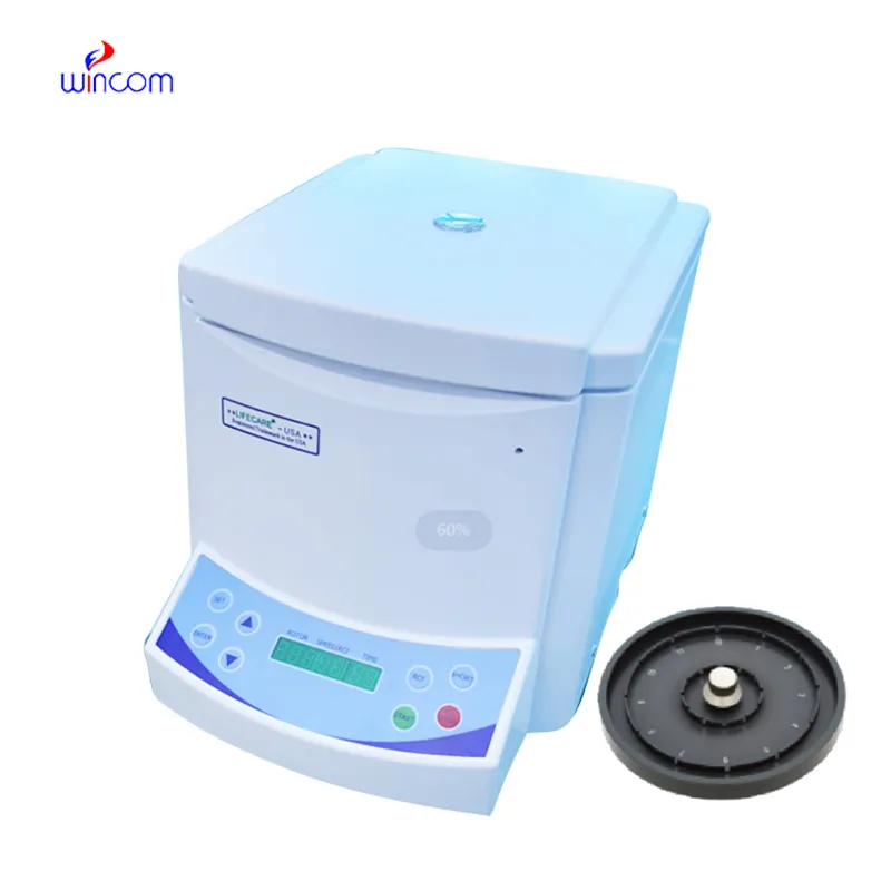
The centrifuge operates quietly and efficiently. It’s compact but surprisingly powerful, making it perfect for daily lab use.
The delivery bed is well-designed and reliable. Our staff finds it simple to operate, and patients feel comfortable using it.
To protect the privacy of our buyers, only public service email domains like Gmail, Yahoo, and MSN will be displayed. Additionally, only a limited portion of the inquiry content will be shown.
Hello, I’m interested in your water bath for laboratory applications. Can you confirm the temperat...
We’re interested in your delivery bed for our maternity department. Please send detailed specifica...
E-mail: [email protected]
Tel: +86-731-84176622
+86-731-84136655
Address: Rm.1507,Xinsancheng Plaza. No.58, Renmin Road(E),Changsha,Hunan,China

 af
af
 es
es
 ar
ar
 tr
tr
 sw
sw
 pt
pt
 th
th
 ur
ur
 bn
bn
 ne
ne
 vi
vi
 km
km
 lo
lo
 de
de
 ru
ru
 fi
fi
 nl
nl
 fa
fa
 fr
fr
 ko
ko