The Electronic Balance is employed by laboratory and hospital personnel for very precise measurement of chemical substances, powders, and living organisms. Its accuracy to the tune of Electronic Balance allows making medications in exact doses, calibrating standards, and preparing diagnostic reagents. Electronic Balance not only gives undistorted and sensitive measures but also improves lab operation, helps to control quality, and keeps clinical research intact. Its importance is appreciated in both hospital tests and medical product development as dependable.

Electronic Balance are used in clinical laboratories for making calibration references that are then checked on the analytics machines. Exact weighing guarantees that the calibration materials keep the same mass values throughout the uses they are subjected to. This application endorses instrument accuracy checks, routine audits in laboratories, and compliance with government regulations. By being a part of trustworthy calibration workflows, Electronic Balance is a major factor and, thus, a contributor to measurement accuracy kept in hospitals' diagnostic devices.

At the medical institutions that are research-driven, Electronic Balance will change to facilitate the analytical methods with higher sensitivity that are in the pipe. The future might bring along the possibility of ultra-low mass samples being accurately measured in molecular diagnostics and sophisticated drug research. This turning development will not only enlarge the experimental capacities of hospital-based research labs but also open new fronts in medical innovation through analytics.

The maintenance of Electronic Balance involves the aspects of storage and inactivity care that come first. The balance should be protected from dust and vibration when it is not in active use. Periodically checking the operational status during long storage prevents unnoticed performance drift. These practices guarantee that Electronic Balance is still capable of accurate use in laboratories, medical and hospital settings.
The precision of Electronic Balance is achieved only in a very controlled environment, which implies regulation of temperature, humidity, and vibration to a minimum level. These parameters are continuously monitored by laboratory technicians to avoid any errors in measurements. Electronic Balance technique provides highly accurate weighing of tiny samples in severe conditions, thus supporting laboratory experiments and hospital-grade analyses of sensitive tests or research that demands careful sample handling.
Q: What maintenance does an Analytical Balance require? A: A periodic cleaning, checking of the calibration, and also verifying the performance are all necessary. Q: Can an Analytical Balance handle continuous daily use? A: Yes, provided that the correct laboratory conditions and rules are followed. Q: Why is leveling important for an Analytical Balance? A: The accuracy and repeatability of the measurements depend on proper leveling. Q: Can Analytical Balances be connected to laboratory systems? A: Most of the models allow connectivity with laboratory information systems. Q: Are Analytical Balances sensitive to vibration? A: Yes, stable weight readings can be disturbed by vibrations.
The water bath performs consistently and maintains a stable temperature even during long experiments. It’s reliable and easy to operate.
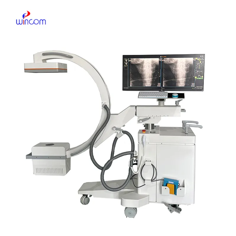
This x-ray machine is reliable and easy to operate. Our technicians appreciate how quickly it processes scans, saving valuable time during busy patient hours.
To protect the privacy of our buyers, only public service email domains like Gmail, Yahoo, and MSN will be displayed. Additionally, only a limited portion of the inquiry content will be shown.
We’re looking for a reliable centrifuge for clinical testing. Can you share the technical specific...
Hello, I’m interested in your water bath for laboratory applications. Can you confirm the temperat...
E-mail: [email protected]
Tel: +86-731-84176622
+86-731-84136655
Address: Rm.1507,Xinsancheng Plaza. No.58, Renmin Road(E),Changsha,Hunan,China

 af
af
 es
es
 ar
ar
 tr
tr
 sw
sw
 pt
pt
 th
th
 ur
ur
 bn
bn
 ne
ne
 vi
vi
 km
km
 lo
lo
 de
de
 ru
ru
 fi
fi
 nl
nl
 fa
fa
 fr
fr
 ko
ko