With the cutting-edge imaging processors, the doppler fetal monitor nearby facilitates real-time, high-resolution images that are paramount in the detection of subtle physiological changes by clinicians. The display is user-friendly for easy parameter modification as well as image marking. The doppler fetal monitor nearby exhibits the mix of effectiveness, mobility, and reliability for a huge range of diagnostic procedures.

The doppler fetal monitor nearby is a tool that medical professionals are using in different departments like pediatrics for the measurement of the size of the organs and neurology for appreciating the anatomy of the soft tissues. It makes it easy for anesthesiologists to perform nerve blocks and vascular access procedures. The doppler fetal monitor nearby increase the speed and the trust of the diagnosis during both routine checkups and highly technical interventions.

Through continued innovations in digital technology, the doppler fetal monitor nearby can be expected to improve and extend its applications within preventive medicine and telemedicine. The next generation of such technologies will facilitate collaboration among experts in real-time using cloud-imaging solutions. The doppler fetal monitor nearby can also work within wearables that include biosensors.

The doppler fetal monitor nearby require a certain set of procedures when it comes to handling them. The images on the display should always be cleaned with soft, lint-free cloths. The probe membranes should always be checked for the absence of cracks. The doppler fetal monitor nearby also require calibration verification.
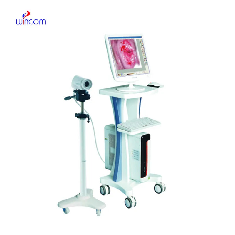
With the advanced imaging technology, the doppler fetal monitor nearby provides physicians unobstructed and precise images of internal organs. It is employed in the early disease diagnosis as well as in patient tracking. The doppler fetal monitor nearby functions by utilizing sound wave reflections to generate dynamic images, qualifying it as an essential tool in modern medical diagnostics. Through the doppler fetal monitor nearby, fast, non-invasive testing is facilitated for real-time assessment to support clinical decisions.
Q: What are the main maintenance requirements for the ultrasound scannert? A: Regular cleaning, proper probe handling, and scheduled inspections help maintain optimal performance. Q: How often should the ultrasound scannert be calibrated? A: Calibration frequency depends on usage levels, but periodic professional checks are recommended. Q: Is the ultrasound scannert suitable for pediatric use? A: Yes, it provides gentle, non-invasive imaging ideal for neonatal and pediatric diagnostics. Q: Does the ultrasound scannert support wireless connectivity? A: Many models include Wi-Fi or Bluetooth features for data sharing and device integration. Q: What materials are used in the ultrasound scannert construction? A: It is built with durable medical-grade components designed to withstand continuous clinical use.
We’ve been using this mri machine for several months, and the image clarity is excellent. It’s reliable and easy for our team to operate.
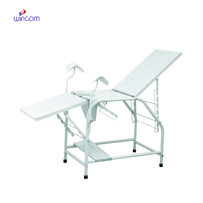
The hospital bed is well-designed and very practical. Patients find it comfortable, and nurses appreciate how simple it is to operate.
To protect the privacy of our buyers, only public service email domains like Gmail, Yahoo, and MSN will be displayed. Additionally, only a limited portion of the inquiry content will be shown.
I’m looking to purchase several microscopes for a research lab. Please let me know the price list ...
We’re currently sourcing an ultrasound scanner for hospital use. Please send product specification...
E-mail: [email protected]
Tel: +86-731-84176622
+86-731-84136655
Address: Rm.1507,Xinsancheng Plaza. No.58, Renmin Road(E),Changsha,Hunan,China

 af
af
 es
es
 ar
ar
 tr
tr
 sw
sw
 pt
pt
 th
th
 ur
ur
 bn
bn
 ne
ne
 vi
vi
 km
km
 lo
lo
 de
de
 ru
ru
 fi
fi
 nl
nl
 fa
fa
 fr
fr
 ko
ko